early detection saves lives
Cardiac imaging that informs action
Modern multimodality cardiac imaging integrates structural and functional assessment to:
- Refine risk stratification
- Support diagnosis
- Clarify disease severity
- Inform management decisions within the appropriate clinical context. 1-3
1. European Society of Cardiology. ESC guidelines for the management of acute coronary syndromes. Published 2023. Accessed 16 December 2025. https://www.escardio.org/Guidelines/Clinical-Practice-Guidelines/Acute-Coronary-Syndromes-ACS-Guidelines
2. Gulati M, Levy PD, Mukherjee D, et al. 2021 AHA/ACC guideline for the evaluation and diagnosis of chest pain. J Am Coll Cardiol. 2021;78(22):e187–e285. doi:10.1016/j.jacc.2021.07.053
3. Storey P. Cardiac imaging: 2022 update. Aust J Gen Pract. 2022;51(9):673–680.
Cardiac Imaging Expertise
Sunshine Coast Radiology offers:
-
Convenient access to CT, ultrasound, nuclear medicine and PET-CT scans
-
Subspeciality cardiac radiologists, expert nuclear medicine physicians and supporting cardiologists that deliver detailed reports and perform interventional procedures
-
A dedicated hotline for referrer support - 07 5430 3926 to:
- Arrange urgent appointments
- Access urgent results
- Speak with a Radiologist
Contact your Referrer Relationship Specialists:
Bernie on 0477 444 230 | brushton@scradiology.com.au
Tremain on 0418 289 686 | tremain@scradiology.com.au
indications & Diagnosing
Cardiac imaging overview
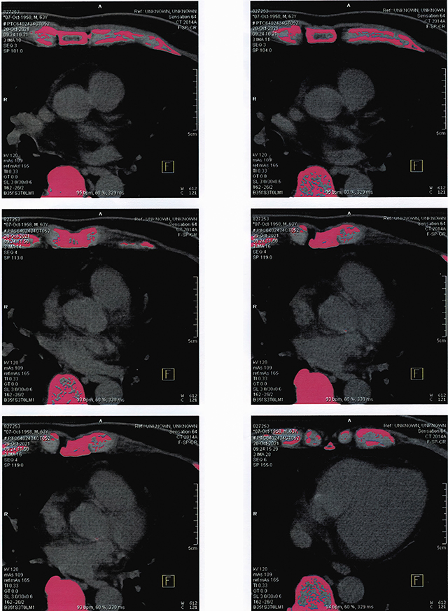
Indications
- Low dose CT to measure coronary calcification and provides a calcium score (Agatston Score)
Best for Diagnosing
- Asymptomatic, intermediate risk patients
Avoid/Consider Alternatives
- Symptomatic chest pain – refer for CTCA instead
- Known CAD
- Stents or bypass grafts
- Atrial fibrillation (less reliable)
Indications
- Coronary anatomy, plaque, stenosis
- Excellent rule-out test
- Shows early development of plaque
- High negative predictive value
- Equivocal stress echocardiogram
Best for Diagnosing
- Chest pain workup
- Low–moderate risk CAD
- Strong family history
- Pre-operative cardiac risk assessment
Avoid/Consider Alternatives
- Atrial fibrillation (affects gating)
- BMI greater than 40kg/m2
- Renal impairment or contrast allergy
- Known high calcium score greater than 1000 AU
Indications
- Assessment of heart and ventricular function, valves, chambers size, and pressures
- Regurgitation/ stenosis grading
Best for Diagnosing
- Symptoms of cardiac ischaemia
- Suspected ventricular hypertrophy or dysfunction
- Pulmonary hypertension
- Valvular, pericardial or embolic disease
- Congenital heart disease
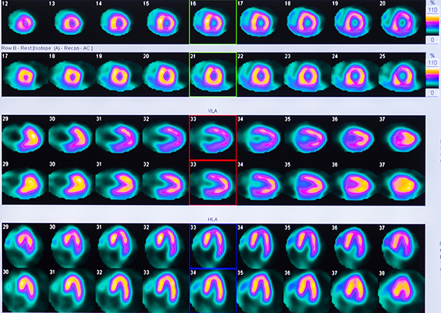
Indications
- Perfusion, ischaemia, viability
Best for Diagnosing
- Complex CAD
- Prior revascularisation
- LBBB
- Unable to exercise adequately
- Equivocal stress echo
cardiac symptoms
Imaging reference guide
Use this general reference guide to help select suitable cardiac imaging when your clinical assessment suggests a possible underlying cardiac cause.
Immediately refer to Emergency Department for cresting troponins, ongoing chest pain, syncope with red flags, or arrhythmia with haemodynamic compromise.
symptom
Recommended Initial Scan
- CT Coronary Angiography +/- CT
- CT Calcium Score
- Echocardiogram
- Alternative Scan:
Stress Echocardiogram if CT
not suitable
Recommended Initial Scan
- Echocardiogram
- Alternative Scan: Diastolic
stress Echocardiogram
Recommended Initial Scan
- Holter Monitor (24-72 hours)
- Event monitor or Holter Monitor (7-14 days) if symptoms are intermittent
Recommended Initial Scan
- ECG + Holter Monitor
- Alternative Scan: Echocardiogram for suspected structural disease
Recommended Follow-up Scan
- Echocardiogram for ongoing symptoms
Recommended Initial Scan
- Echocardiogram
Recommended Initial Scan
- ECG
Recommended Follow up Scan
- Echocardiogram if clinically indicated
Further Follow-up Scan
- Holter Monitor if clinically indicated
Recommended Initial Scan
- CT Coronary Angiography + Stress Echocardiogram
Recommended Follow-up Scan
- Myocardial Perfusion Scan if uncertainty persists
Recommended Initial Scan
- Cardiac MRI